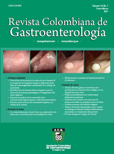
La cromoendoscopia virtual i-Scan y su aplicación en la detección y caracterización de los pólipos de colon
DOI:
https://doi.org/10.22516/25007440.127Palabras clave:
i-Scan, colonoscopia, pólipoResumen
La limitación de la colonoscopia con luz blanca respecto a la omisión de neoplasias avanzadas en un 6%, y de adenomas hasta un 25%, ha motivado el desarrollo de técnicas como la cromoendoscopia virtual, entre ellas el sistema i-Scan, para detectar un mayor número de pólipos, como estrategia de prevención del cáncer de colon. Con esta revisión se resume la aplicación actual de este método en la detección de adenomas y su caracterización. Se ha encontrado que este nuevo sistema es una alternativa posible en nuestro medio a las ya existentes y mejor conocidas, como el NBI y FICE.
Descargas
Referencias bibliográficas
Winawer SJ, Zauber AG, Ho MN, et al. Prevention of colorectal cancer by colonoscopy polypectomy. The National Polyp Study Workgroup. N Engl J Med. 1993;329:1977-81. doi: https://doi.org/10.1056/NEJM199312303292701
Winawer SJ, Zauber AG, O'Brien MJ, et al. Randomized comparison of surveillance intervals after colonoscopic removal of newly diagnosed adenomatous polyps. N Engl J Med. 1993;328:901-6. doi: https://doi.org/10.1056/NEJM199304013281301
Bressler B, Paszat LF, Chen Z, et al. Rates of new or missed colorectal cancers after colonoscopy and their risk factors: a population-based analysis. Gastroenterology. 2007;132:96-112. doi: https://doi.org/10.1053/j.gastro.2006.10.027
Van Rijn JC, Eitsma JB, Stoker J, et al. Polyp miss rate determined by tandem colonoscopy: a systematic review. Am J Gastroenterol. 2006;101:343-50. doi: https://doi.org/10.1111/j.1572-0241.2006.00390.x
Rex DK, Cutler CS, Lemmel GT, et al. Colonoscopic miss rates of adenomas determined by back to back colonoscopies. Gastroenterology. 2007;132:96-102.
Devuni D, Vaziri H, Anderson J. Chromocolonoscopy. Gastroenterol Clin N Am. 2013;42:521-45. doi: https://doi.org/10.1016/j.gtc.2013.05.002
Kudo S, Hirota S, Nakajima T, et al. Colorectal tumours and Pit Pattern. J Clin Pathol. 1994;47:880-5. doi: https://doi.org/10.1136/jcp.47.10.880
Tischendorf J, Wasmuth HE, Koch A, et al. Value of magnifying chromoendoscopy and narrow band imaging (NBI) in classifying colorectal polyps a prospective controlled study. Endoscopy. 2007;39:1092-6. doi: https://doi.org/10.1055/s-2007-966781
Huang Q, Fukami N, Kashida H, et al. Interobserver and Intraobserver consistency in the endoscopic assessment of colon pit patterns. Gastrointest Endosc. 2001;60:520-6. doi: https://doi.org/10.1016/S0016-5107(04)01880-2
Lopez- Cerón L, Sanabria E, Pellise M. Colonic polyps is it useful to characterize them with advance endoscopy? World J Gastroenterol. 2014;26:8449-57.
Neuman H, Fujishro M, Wilcox M, et al. Present and future perspectives of virtual chromoendoscopy with i-scan and optical enhancement technology. Dig Endosc. 2014;26(Suppl 1):43-51. doi: https://doi.org/10.1111/den.12190
Neuman H, Nagel A, Buda A. Advance endoscopic imaging to improve adenoma detection. World J Gastrointest Endosc. 2015;7:224-9. doi: https://doi.org/10.4253/wjge.v7.i3.224
Reyes-Dorantes A. Nuevas técnicas de imagen (i-Scan, NBI, FICE). Rev Gastroenterol Mex. 2011;76:(Suppl 1):134-6.
Yeung TM, Mortesen NJ. Advances in endoscopic visualization of colorectal polyps. Colorectal Dis. 2011;13:352-9. doi: https://doi.org/10.1111/j.1463-1318.2009.02142.x
Galloro G. High technology imaging in digestive endoscopy. World J Gastrointest Endosc. 2012;16;4(2):22-7.
Masci E, Mangiavillano B, Costa C, et al. Interobserver agreement among endoscopists on evaluation of polypoid colorectal lesions visualized with Pentax i-Scan Technique. Dig Liver Dis. 2013;45:207-10. doi: https://doi.org/10.1016/j.dld.2012.09.012
Banks M, Haidry R, Butt M, et al. High resolution colonoscopy in a bowel cancer screening program improves polyp detection. World J Gastroenterol. 2011;17:4308-13. doi: https://doi.org/10.3748/wjg.v17.i38.4308
Kwon RS, Adler DG, Chand B, et al. High-resolution and High-magnification endoscopes. Gastrointetinal Endosc. 2009;69:399-407. doi: https://doi.org/10.1016/j.gie.2008.12.049
Ventakaraman S, Ragunath K. Advance endoscopic imaging: a review of commercially aviable technologies. Clin Gastroenterol Hepatol. 2014;12:368-76. doi: https://doi.org/10.1016/j.cgh.2013.06.015
Hancock S, Bowman E, Prabakaran J, et al. Use of i-scan endoscopic image enhancement technology and therapeutic endoscopy: a case series and review of the literature. Diagn Ther Endosc. 2012;2012:193570. doi: https://doi.org/10.1155/2012/193570
Kodashima S, Fujishiro M. Novel image-enhanced endoscopy with i-scan technology. World J Gastroenterol. 2010;16:1043-9. doi: https://doi.org/10.3748/wjg.v16.i9.1043
Cho JH. Advanced imaging technology other than narrow band imaging. Clin Endosc. 2015:48:503-10. doi: https://doi.org/10.5946/ce.2015.48.6.503
Hoffman A, Sar F, Goetz M, et al. High definition colonoscopy combined with i-scan is superior in the detection of colorectal neoplasias compared with standard videocolonoscopy: a prospective randomized controlled trial. Endoscopy. 2010;47:827-33. doi: https://doi.org/10.1055/s-0030-1255713
Hoffman A, Loth L, Rey JW, et al. High definition plus colonoscopy combined with i-scan tone enhancement vs. high definition colonoscopy for colorectal neoplasia: A randomized trial. Dig Liver Dis. 2014;46:991-6. doi: https://doi.org/10.1016/j.dld.2014.07.169
Bowman E, Pfau P, Mitra A, et al. High definition colonoscopy combined with i-SCAN imaging technology is superior in the detection of adenomas and advanced lessions compared to high definition colonoscopy alone. Diagn Ther Endosc. 2015;2015:167406. doi: https://doi.org/10.1155/2015/167406
Hong SN, Choe WH, Lee JH, et al. Prospective, randomized, back-to-back trial evaluating the usefulness of i-SCAN in screening colonoscopy. Gastrointest Endosc. 2012;75(5):1011-21. doi: https://doi.org/10.1016/j.gie.2011.11.040
Hoffman A, Kagel C, Goetz M, et al. Recognition and characterization of small colonic neoplasia with-definition colonoscopy using i-scan as precise chromoendoscopy. Dig Liver Dis. 2009;42;45-50. doi: https://doi.org/10.1016/j.dld.2009.04.005
Pigo F, Bertani H, Barbera C, et al. I-Scan high-definition white light endoscopy and colorectal polyps: prediction of histology interobserver and intraobserver agreement. Int J Colorectal Dis. 2013;28:399-406. doi: https://doi.org/10.1007/s00384-012-1583-7
Lee Ch, Lee SH, Hwagbo Y. Narrow-band imaging versus I-Scan for the real time histological prediction of diminutive colonic polyps: a prospective comparative study by using the simple unified endoscopic classification. Gastrointest Endosc. 2011;74:603-9. doi: https://doi.org/10.1016/j.gie.2011.04.049
Hong SN, Choe WC, Lee JH, et al. Prospective, randomized, back-to-back trial evaluating the usefulness of i-SCAN screening colonoscopy. Gastrointest Endosc. 2012;75:1011-21. doi: https://doi.org/10.1016/j.gie.2011.11.040
Wanders L, East J, Uitentius S, et al. Diagnostic performance of narrowed a spectrum endoscopy autofluorescense imaging, and confocal laser endomicroscopy for optical diagnosis of colonic polyps: a meta-analysis. Lancet. 2013;14:1337-47. doi: https://doi.org/10.1016/S1470-2045(13)70509-6
Bouwens M, Ridder R, Masclee A, et al. Optical diagnosis of colorectal polyps using high-definition i-scan: an educational experience. World J Gastroenterol. 2013;19:4334-43. doi: https://doi.org/10.3748/wjg.v19.i27.4334
Neuman H, Vieth M, Fry L, et al. Learning curve of virtual chromo endoscopy for the prediction of hyperplastic and adenomatous colorectal lesions: a prospective 2-center study. Gastrointest Endosc. 2013;78:115-20. doi: https://doi.org/10.1016/j.gie.2013.02.001
Rex DK, Kahi C, O´Brien M, et al. The American Society for Gastrointestinal Endoscopy PIVI (Preservation and Incorporation of Valuable Endoscopic Innovations) on real-time endoscopic assessment of the histology of diminute colorectal polyps. Gastrointest Endosc. 2011;73:419-22. doi: https://doi.org/10.1016/j.gie.2011.01.023
Kaltenbach T, Rex D, Wilson A, et al. Implementation of optical diagnosis for colorectal polyps: standardization of studies is needed. Clin Gastroenterol Hepatol. 2015;13:6-10. doi: https://doi.org/10.1016/j.cgh.2014.10.009
Hassan C, Repici A, Zullo A, et al. New paradigms for colonoscopic management of diminutive colorectal polyps: predict, resect, and discard or do not resect? Clin Endosc. 2013;48:130-7. doi: https://doi.org/10.5946/ce.2013.46.2.130
Abu Dayyeh BK, Thosani N, Konda V, et al. ASGE Technology Committee systematic review and meta-analysis assessing the ASGE PIVI thresholds for adopting real-time endoscopic assessment of the histology of diminutive colorectal polyps. Gastrointest Endosc. 2015;81:502-16. doi: https://doi.org/10.1016/j.gie.2014.12.022
Basford P, Longcroft-Wheaton G, Higgins B, et al. High-definition endoscopy with i-Scan for evaluation of small colon polyps: the HisCOPE study. Gastrointest Endosc. 2014;79:111-8. doi: https://doi.org/10.1016/j.gie.2013.06.013
Wilson BA. Optical diagnosis of small colorectal polyps during colonoscopy: When to resect and during colonoscopy: When to resect and discard? Best Pract Res Clin Gastroenterol. 2015;29:639-49. doi: https://doi.org/10.1016/j.bpg.2015.06.007
Gomez V, Wallace M. Advances in diagnostic and therapeutic colonoscopy. Curr Opin Gastroenterol. 2014;30:63-8. doi: https://doi.org/10.1097/MOG.0000000000000026
Kiesslich R. Novel colonoscopic imaging techniques. Gastroenterol Hepatol. 2013;9:241-4.
Kaminski M, Hassan C, Bisschops R, et al. Advance imaging for detection and differentiation of colorectal neoplasia: European Society of Gastrointestinal Endoscopy (ESGE) Guideline. Endoscopy. 2014:46;435-49. doi: https://doi.org/10.1055/s-0034-1365348
Bath Y, Abu Dayyeh B, Chauhan S, et al. High-definition and high-magnification endoscopes. Gastrointest Endosc. 2014;80:919-27. doi: https://doi.org/10.1016/j.gie.2014.06.019
Jang JY. The past, present, and future of image- enhanced. Endoscopy Clin Endosc. 2015;48:466-75. doi: https://doi.org/10.5946/ce.2015.48.6.466
Descargas
Publicado
Cómo citar
Número
Sección
Licencia
Aquellos autores/as que tengan publicaciones con esta revista, aceptan los términos siguientes:
Los autores/as ceden sus derechos de autor y garantizarán a la revista el derecho de primera publicación de su obra, el cuál estará simultáneamente sujeto a la Licencia de reconocimiento de Creative Commons que permite a terceros compartir la obra siempre que se indique su autor y su primera publicación en esta revista.
Los contenidos están protegidos bajo una licencia de Creative Commons Reconocimiento-NoComercial-SinObraDerivada 4.0 Internacional.


