Intestinal Vasculitis and Endoscopic Video Capsule Findings: Case Report and Literature Review
DOI:
https://doi.org/10.22516/25007440.812Keywords:
Vasculitis, Small intestine, Capsule endoscopyAbstract
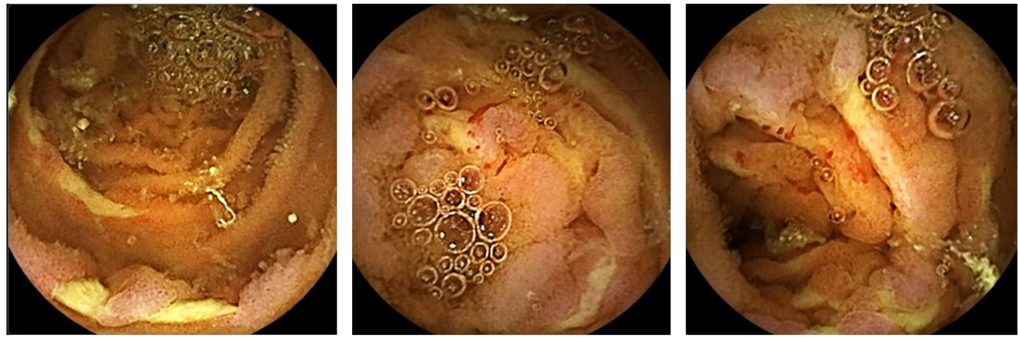
Systemic vasculitis can affect blood vessels of all sizes, causing necrosis and inflammation. Granulomatosis with polyangiitis (GPA) is a vasculitis of small and medium blood vessels. Clinical manifestations may be local or diffuse in the gastrointestinal tract. We present a patient’s case with systemic vasculitis associated with anti-cytoplasmic antibodies (ANCA), myeloperoxidase (MPO) antibodies (microscopic polyangiitis) confirmed through renal biopsy, presenting a 15-day clinical picture consisting of asthenia, adynamia, subjective oliguria, edema of the upper and lower limbs, hyporexia, and melena. The digital rectal examination was positive for melena in the physical examination, later presenting melena with secondary anemization. An endoscopic video capsule was performed, showing findings compatible with enteric vasculitis. During his stay, he presented multisystemic involvement due to renal, pulmonary, neurological, and gastrointestinal involvement, managed in the intensive care unit (ICU), where systemic steroid pulses and hemodialysis started.
Conclusion: although gastrointestinal vasculitis is a rare complication, it occurs and threats patients’ lives. Differential diagnosis should cover other inflammatory diseases, especially Crohn’s disease. Gastrointestinal vasculitis early diagnosis significantly influences prognosis, as prompt steroid therapy can change the course of the disease.
Downloads
References
Pagnoux C, Mahr A, Cohen P, Guillevin L. Presentation and outcome of gastrointestinal involvement in systemic necrotizing vasculitides: analysis of 62 patients with polyarteritis nodosa, microscopic polyangiitis, Wegener granulomatosis, Churg-Strauss syndrome, or rheumatoid arthritis-associated vasculitis. Medicine (Baltimore). 2005;84(2):115-128. https://doi.org/10.1097/01.md.0000158825.87055.0b
Ge ZZ, Hu YB, Xiao SD. Capsule endoscopy and push enteroscopy in the diagnosis of obscure gastrointestinal bleeding. Chin Med J (Engl). 2004;117(7):1045-9.
Proctor DD, Panzini LA. Isolated and diffuse ulcers of the small intestine. En: Feldman M, Friedman LS, Sleisenger MH (editores) Sleisenger and Fordran’s gastrointestinal and liver disease. 7.a edición., Filadelfia: Saunders; 2002. p. 2081-8.
Ghosh S, Watts D, Kinnear M. Management of gastrointestinal haemorrhage. Postgrad Med J. 2002;78(915):4-14. https://doi.org/10.1136/pmj.78.915.4
Yamamoto H, Kita H, Sunada K, Hayashi Y, Sato H, Yano T, et al. Clinical outcomes of double-balloon endoscopy for the diagnosis and treatment of small-intestinal diseases. Clin Gastroenterol Hepatol. 2004;2(11):1010-6. https://doi.org/10.1016/S1542-3565(04)00453-7
Tang SJ, Zanati S, Dubcenco E, Christodoulou D, Cirocco M, Kandel G, Kortan P, Haber GB, Marcon NE. Capsule endoscopy regional transit abnormality: a sign of underlying small bowel pathology. Gastrointest Endosc. 2003;58(4):598-602. https://doi.org/10.1067/S0016-5107(03)01963-1
Liangpunsakul S, Maglinte DD, Rex DK. Comparison of wireless capsule endoscopy and conventional radiologic methods in the diagnosis of small bowel disease. Gastrointest Endosc Clin N Am. 2004;14(1):43-50. https://doi.org/10.1016/j.giec.2003.10.004
Costamagna G, Shah SK, Riccioni ME, Foschia F, Mutignani M, Perri V, Vecchioli A, Brizi MG, Picciocchi A, Marano P. A prospective trial comparing small bowel radiographs and video capsule endoscopy for suspected small bowel disease. Gastroenterology. 2002;123(4):999-1005. https://doi.org/10.1053/gast.2002.35988
Liangpunsakul S, Chadalawada V, Rex DK, Maglinte D, Lappas J. Wireless capsule endoscopy detects small bowel ulcers in patients with normal results from state of the art enteroclysis. Am J Gastroenterol. 2003;98(6):1295-8. https://doi.org/10.1111/j.1572-0241.2003.07471.x
Hara AK, Leighton JA, Sharma VK, Heigh RI, Fleischer DE. Imaging of small bowel disease: comparison of capsule endoscopy, standard endoscopy, barium examination, and CT. Radiographics. 2005;25(3):697-711; discussion 711-8. https://doi.org/10.1148/rg.253045134
Voderholzer WA, Beinhoelzl J, Rogalla P, Murrer S, Schachschal G, Lochs H, et al. Small bowel involvement in Crohn’s disease: a prospective comparison of wireless capsule endoscopy and computed tomography enteroclysis. Gut. 2005;54(3):369-73. https://doi.org/10.1136/gut.2004.040055
Hoffman GS, Kerr GS, Leavitt RY, Hallahan CW, Lebovics RS, Travis WD, et al. Wegener granulomatosis: an analysis of 158 patients. Ann Intern Med. 1992;116(6):488-98. https://doi.org/10.7326/0003-4819-116-6-488
Masiak A, Zdrojewski Ł, Zdrojewski Z, Bułło-Piontecka B, Rutkowski B. Gastrointestinal tract involvement in granulomatosis with polyangiitis. Prz Gastroenterol. 2016;11(4):270-275. https://doi.org/10.5114/pg.2016.57887
Revzin MV, Pellerito JS. Doppler Ultrasound of the Mesenteric Vasculature. En: Pellerito JS, Polak JF (editores). Introduction to Vascular Ultrasonography. 7.a edición. Elsevier; 2019. p. 547-81.
Downloads
Published
How to Cite
Issue
Section
License
Copyright (c) 2022 Revista colombiana de Gastroenterología

This work is licensed under a Creative Commons Attribution-NonCommercial-NoDerivatives 4.0 International License.
Aquellos autores/as que tengan publicaciones con esta revista, aceptan los términos siguientes:
Los autores/as ceden sus derechos de autor y garantizarán a la revista el derecho de primera publicación de su obra, el cuál estará simultáneamente sujeto a la Licencia de reconocimiento de Creative Commons que permite a terceros compartir la obra siempre que se indique su autor y su primera publicación en esta revista.
Los contenidos están protegidos bajo una licencia de Creative Commons Reconocimiento-NoComercial-SinObraDerivada 4.0 Internacional.