Liver Abscess Caused by Enterobius vermicularis as a Differential Diagnosis for Liver Metastasis in Colorectal Cancer, Case Report
DOI:
https://doi.org/10.22516/25007440.790Keywords:
Colon neoplasms, Rectum neoplasms, Liver abscess, EnterobiusAbstract
Introduction: colorectal cancer is the fourth leading cause of cancer-related mortality worldwide. The identification of the metastases of this tumor in the preoperative stage is increasingly frequent due to the imaging studies currently available. We present the case of a patient with an infection caused by Enterobius vermicularis that simulates the presence of liver metastases.
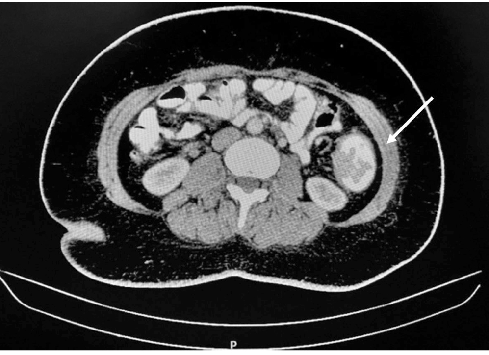
Case presentation: a female patient from a rural area showing a one-year abdominal pain evolution associated with lower gastrointestinal tract bleeding and weight loss. Endoscopic imaging and studies displayed a tumor lesion in the sigmoid colon, with biopsies reporting sigmoid colon adenocarcinoma and liver lesions suggesting malignancy. Anterior resection of the rectum and sigmoid was performed with high anastomosis and liver biopsies, which ruled out malignancy and reported the presence of liver infection by E. vermicularis.
Discussion: in this case, the hepatic E. vermicularis infection was rare. This infection can simulate the presence of liver metastases; therefore, it should be considered a differential diagnosis of metastatic colorectal cancer.
Downloads
References
Bray F, Ferlay J, Soerjomataram I, Siegel RL, Torre LA, Jemal A. Global cancer statistics 2018: GLOBOCAN estimates of incidence and mortality worldwide for 36 cancers in 185 countries. CA Cancer J Clin. 2018;68(6):394-424. https://doi.org/10.3322/caac.21492
Arkoulis N, Zerbinis H, Simatos G, Nisiotis A. Enterobius vermicularis (pinworm) infection of the liver mimicking malignancy: Presentation of a new case and review of current literature. Int J Surg Case Rep. 2012;3(1):6-9. https://doi.org/10.1016/j.ijscr.2011.10.003
Ng WS, Gallagher J, McCaughan G. “Pinworm” infection of the liver: unusual CT appearance leading to hepatic resection. Dig Dis Sci. 2004 Mar;49(3):466-8. https://doi.org/10.1023/b:ddas.0000020505.46611.a0
Valderrama-Treviño AI, Barrera-Mera B, Ceballos-Villalva JC, Montalvo-Javé EE. Hepatic Metastasis from Colorectal Cancer. Euroasian J Hepatogastroenterol. 2017;7(2):166-175. https://doi.org/10.5005/jp-journals-10018-1241
Cook GC. Enterobius vermicularis infection. Gut. 1994;35(9):1159-62. https://doi.org/10.1136/gut.35.9.1159
Little MD, Cuello CJ, D’Alessandro A. Granuloma of the liver due to Enterobius vermicularis. Report of a case. Am J Trop Med Hyg. 1973;22(4):567-9. https://doi.org/10.4269/ajtmh.1973.22.567
Kim HY, Kim CW, Kim DR, Cho YW, Cho JY, Kim WJ, et al. Recurrent pyogenic liver abscess as a presenting manifestation of colorectal cancer. Korean J Intern Med. 2017;32(1):174-177. https://doi.org/10.3904/kjim.2015.301
Meyers W, Neafie R, Marty A, Wear DJ. Pathology of infectious disease, volume I Helminthiases. Amer Registry of Pathology; 2000.
Bhullar DS, Barriuso J, Mullamitha S, Saunders MP, O’Dwyer ST, Aziz O. Biomarker concordance between primary colorectal cancer and its metastases. EBioMedicine. 2019;40:363-374. https://doi.org/10.1016/j.ebiom.2019.01.050
Chow FC, Chok KS. Colorectal liver metastases: An update on multidisciplinary approach. World J Hepatol. 2019;11(2):150-172. https://doi.org/10.4254/wjh.v11.i2.150
Oh JG, Choi SY, Lee MH, Lee JE, Yi BH, Kim SS, et al. Differentiation of hepatic abscess from metastasis on contrast-enhanced dynamic computed tomography in patients with a history of extrahepatic malignancy: emphasis on dynamic change of arterial rim enhancement. Abdom Radiol (NY). 2019;44(2):529-538. https://doi.org/10.1007/s00261-018-1766-y
Tirumani SH, Kim KW, Nishino M, Howard SA, Krajewski KM, Jagannathan JP, et al. Update on the role of imaging in management of metastatic colorectal cancer. Radiographics. 2014;34(7):1908-28. https://doi.org/10.1148/rg.347130090

Downloads
Published
How to Cite
Issue
Section
License
Copyright (c) 2022 Revista colombiana de Gastroenterología

This work is licensed under a Creative Commons Attribution-NonCommercial-NoDerivatives 4.0 International License.
Aquellos autores/as que tengan publicaciones con esta revista, aceptan los términos siguientes:
Los autores/as ceden sus derechos de autor y garantizarán a la revista el derecho de primera publicación de su obra, el cuál estará simultáneamente sujeto a la Licencia de reconocimiento de Creative Commons que permite a terceros compartir la obra siempre que se indique su autor y su primera publicación en esta revista.
Los contenidos están protegidos bajo una licencia de Creative Commons Reconocimiento-NoComercial-SinObraDerivada 4.0 Internacional.


