Dieulafoy’s Lesion: A Case Report with an Atypical Anatomical Variation
DOI:
https://doi.org/10.22516/25007440.1176Keywords:
Dieulafoy’s lesion, Gastrointestinal Bleeding, Angiodysplasia, Hemorrhagic shockAbstract
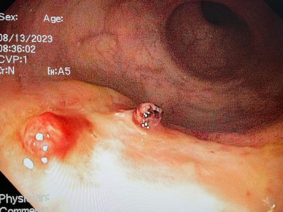
Dieulafoy’s lesion is a rare and potentially life-threatening cause of gastrointestinal bleeding, resulting from vascular injury due to submucosal erosion that penetrates through the mucosa. It can lead to gastrointestinal hemorrhage with severe complications. This condition accounts for 1%–2% of gastrointestinal bleeding cases, most commonly occurring in the gastric fundus, and it typically affects individuals over 50 years of age with multiple comorbidities. This report describes a rare etiology of gastrointestinal bleeding associated with a thrombectomy and an atypical anatomical location.
Downloads
References
Juler GL, Labitzke HG, Lamb R, Allen R. The pathogenesis of Dieulafoy’s gastric erosion. Am J Gastroenterol. 1984;79(3):195-200.
Nojkov B, Cappell MS. Gastrointestinal bleeding from Dieulafoy’s lesion: Clinical presentation, endoscopic findings, and endoscopic therapy. World J Gastrointest Endosc. 2015;7(4):295-307. https://doi.org/10.4253/wjge.v7.i4.295
Baxter M, Aly EH. Dieulafoy’s lesion: current trends in diagnosis and management. Ann R Coll Surg Engl. 2010;92(7):548-54. https://doi.org/10.1308/003588410X12699663905311
Batouli A, Kazemi A, Hartman MS, Heller MT, Midian R, Lupetin AR. Dieulafoy lesion: CT diagnosis of this lesser-known cause of gastrointestinal bleeding. Clin Radiol. 2015;70(6):661-6. https://doi.org/10.1016/j.crad.2015.02.005
Kalman DR, Banner BF, Barnard GF. Rectal Dieulafoy’s or angiodysplasia? Gastrointest Endosc. 1997;46(1):91-2. https://doi.org/10.1016/S0016-5107(97)70224-4
Hokama A, Takeshima Y, Toyoda A, Yonamine Y, Tomiyama R, Kinjo F, et al. Images of interest. Gastrointestinal: rectal Dieulafoy lesion. J Gastroenterol Hepatol. 2005;20(8):1303. https://doi.org/10.1111/j.1440-1746.2005.04056.x
Goldkamp W, Goldberg R, Patel K, Tombazzi C. Rare Case of GI Bleeding: Rectal Dieulafoy’s Lesion. Am J Gastroenterol. 2014;109:S414. https://doi.org/10.14309/00000434-201410002-01397
Dy NM, Gostout CJ, Balm RK. Bleeding from the endoscopically-identified Dieulafoy lesion of the proximal small intestine and colon. Am J Gastroenterol. 1995;90(1):108-11.
Reilly HF, Al-Kawas FH. Dieulafoy’s lesion. Diagnosis and management. Dig Dis Sci. 1991;36(12):1702-7. https://doi.org/10.1007/BF01296613
Inayat F, Hussain A, Yahya S, Weissman S, Sarfraz N, Faisal MS, et al. Rectal Dieulafoy’s lesion: a comprehensive review of patient characteristics, presentation patterns, diagnosis, management, and clinical outcomes. Transl Gastroenterol Hepatol. 2022;7:10. https://doi.org/10.21037/tgh.2020.02.17
Yamaguchi Y, Yamato T, Katsumi N, Imao Y, Aoki K, Morita Y, et al. Short-term and long-term benefits of endoscopic hemoclip application for Dieulafoy’s lesion in the upper GI tract. Gastrointest Endosc. 2003;57(6): 653-6. https://doi.org/10.1067/mge.2003.183
Kaneko M, Nozawa H, Tsuji Y, Emoto S, Murono K, Nishikawa T, et al. Multidetector-Row Computed Tomography and Colonoscopy for Detecting a Rectal Dieulafoy Lesion as a Source of Lower Gastrointestinal Hemorrhage. Case Rep Gastroenterol. 2018;12(1):202-206. https://doi.org/10.1159/000488973
Eisenberg D, Bell R. Intraoperative endoscopy: a requisite tool for laparoscopic resection of unusual gastrointestinal lesions--a case series. J Surg Res. 2009;155(2):318-20. https://doi.org/10.1016/j.jss.2008.06.046
Downloads
Published
How to Cite
Issue
Section
License
Copyright (c) 2024 Revista colombiana de Gastroenterología

This work is licensed under a Creative Commons Attribution-NonCommercial-NoDerivatives 4.0 International License.
Aquellos autores/as que tengan publicaciones con esta revista, aceptan los términos siguientes:
Los autores/as ceden sus derechos de autor y garantizarán a la revista el derecho de primera publicación de su obra, el cuál estará simultáneamente sujeto a la Licencia de reconocimiento de Creative Commons que permite a terceros compartir la obra siempre que se indique su autor y su primera publicación en esta revista.
Los contenidos están protegidos bajo una licencia de Creative Commons Reconocimiento-NoComercial-SinObraDerivada 4.0 Internacional.