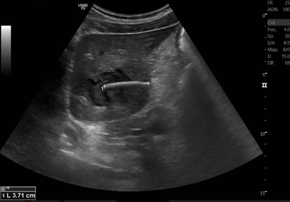
Absceso hepático por espina de pescado: reporte de caso
DOI:
https://doi.org/10.22516/25007440.1117Palabras clave:
Cirugía general, hepatología, reacción a cuerpo extrañoResumen
La presencia de cuerpos extraños en el hígado es una entidad infrecuente. Se reporta el caso de un paciente con absceso hepático por espina de pescado secundario a la migración de esta desde el tracto gastrointestinal. Las manifestaciones clínicas son inespecíficas; por eso, en este caso, el diagnóstico oportuno y el abordaje quirúrgico precoz permitieron disminuir la estancia hospitalaria, el riesgo de complicaciones y la muerte.
Descargas
Referencias bibliográficas
Yu Y, Guo L, Hu C, Chen K. Spectral CT imaging in the differential diagnosis of necrotic hepatocellular carcinoma and hepatic abscess. Clin Radiol. 2014;69(12):e517-e524. https://doi.org/10.1016/j.crad.2014.08.018
Chen CH, Wu SS, Chang HC, Chang YJ. Initial presentations and final outcomes of primary pyogenic liver abscess: a cross-sectional study. BMC Gastroenterol. 2014;14:133. https://doi.org/10.1186/1471-230X-14-133
Lin ACM, Yeh DY, Hsu YH et al. Diagnosis of pyogenic liver abscess by abdominal ultrasonography in the emergency department. Emerg Med J. 2009;26(4):273-275. https://doi.org/10.1136/emj.2007.049254
Mavilia MG, Molina M, Wu GY. The Evolving Nature of Hepatic Abscess: A Review. J Clin Transl Hepatol. 2016;4(2):158-68. https://doi.org/10.14218/JCTH.2016.00004
Lodhi S, Sarwari AR, Muzammil M, Salam A, Smego RA. Features distinguishing amoebic from pyogenic liver abscess: a review of 577 adult cases. Trop Med Int Health. 2004;9(6):718-23. https://doi.org/10.1111/j.1365-3156.2004.01246.x
López MC, Quiroz DA, Pinilla AE. Diagnóstico de amebiasis intestinal y extraintestinal [Internet]. Acta Med Colomb. 2008;33(2):75-83 [consultado el 16 de septiembre de 2024]. Disponible en: https://www.actamedicacolombiana.com/ojs/index.php/actamed/article/view/1759
MacFadden DR, Penner TP, Gold WL. Persistent epigastric pain in an 80-year-old man. CMAJ. 2011;183(8):925-8. https://doi.org/10.1503/cmaj.101510
Mohanty S, Panigrahi MK, Turuk J, Dhal S. Liver Abscess due to Streptococcus constellatus in an Immunocompetent Adult: A Less Known Entity. J Natl Med Assoc. 2018;110(6):591-595. https://doi.org/10.1016/j.jnma.2018.03.006
Tsui BC, Mossey J: Occult liver abscess following clinically unsuspected ingestion of foreign bodies. Can J Gastroenterol. 1997;11(5):445-8. https://doi.org/10.1155/1997/815876
Bandeira-de-Mello RG, Bondar G, Schneider E, Wiener-Stensmann IC, Gressler JB, Kruel CRP. Pyogenic liver abscess secondary to foreign body (fish bone) treated by laparoscopy: A case report. Ann Hepatol. 2018;17(1):169-73. https://doi.org/10.5604/01.3001.0010.7550
Santos SA, Alberto SC, Cruz E, Pires E, Figueira T, Coimbra E, et al. Hepatic abscess induced by foreign body: case report and literature review. World J Gastroenterol. 2007;13(9):1466-70. https://doi.org/10.3748/wjg.v13.i9.1466
Lambert A. Abscess of the liver of unusual origin. NY Med J. 1898:177-78.
Chintamani, Singhal V, Lubhana P, Durkhere R, Bhandari S. Liver abscess secondary to a broken needle migration--a case report. BMC Surg. 2003;3:8. https://doi.org/10.1186/1471-2482-3-8
Hunter TB, Taljanovic MS. Foreign bodies. Radiographics. 2003;23(3):731-57. https://doi.org/10.1148/rg.233025137
Horii K, Yamazaki O, Matsuyama M, Higaki I, Kawai S, Sakaue Y. Successful treatment of a hepatic abscess that formed secondary to fish bone penetration by percutaneous transhepatic removal of the foreign body: report of a case. Surg Today 1999;29(9):922-926. https://doi.org/10.1007/BF02482788
Ndong A, Tendeng JN, Ndoye NA, Diao ML, Dieye A, Diallo AC, et al. Predictive risk factors for liver abscess rupture: A prospective study of 138 cases. Arch Clin Gastroenterol. 2020;6(1):1-5. https://doi.org/10.17352/2455-2283.000067
Webb WA. Management of foreign bodies of the upper gastrointestinal tract: update. Gastrointest Endosc. 1995;41(1):39-51. https://doi.org/10.1016/s0016-5107(95)70274-1
Reid-Lombardo KM, Khan S, Sclabas G. Hepatic cysts and liver abscess. Surg Clin North Am. 2010;90(4):679-97. https://doi.org/10.1016/j.suc.2010.04.004
Skjoldbye B, Brahe NE, Jess P, Nolsøe CP. Laparoskopisk ultralydskanning af lever, galdeveje og pancreas med styrbart lydhoved. Ugeskr Laeger. 1995;157(5):580-3.
Foley EF, Kolecki RV, Schirmer BD. The accuracy of laparoscopic ultrasound in the detection of colorectal cancer liver metastases. Am J Surg 1998;176(3):262-264. https://doi.org/10.1016/S0002-9610(98)00147-0
Salmon M, Doniger SJ. Ingested foreign bodies: A case series demonstrating a novel application of point-of-care ultrasonography in children. Pediatr Emerg Care. 2013;29(7):870-873. https://doi.org/10.1097/PEC.0b013e3182999ba3

Descargas
Publicado
Cómo citar
Número
Sección
Licencia
Derechos de autor 2024 Revista colombiana de Gastroenterología

Esta obra está bajo una licencia internacional Creative Commons Atribución-NoComercial-SinDerivadas 4.0.
Aquellos autores/as que tengan publicaciones con esta revista, aceptan los términos siguientes:
Los autores/as ceden sus derechos de autor y garantizarán a la revista el derecho de primera publicación de su obra, el cuál estará simultáneamente sujeto a la Licencia de reconocimiento de Creative Commons que permite a terceros compartir la obra siempre que se indique su autor y su primera publicación en esta revista.
Los contenidos están protegidos bajo una licencia de Creative Commons Reconocimiento-NoComercial-SinObraDerivada 4.0 Internacional.


